


Diagnostic Imaging | MRI
MRI
Magnetic Resonance Imaging (MRI) is primarily a medical imaging technique most commonly used in radiology to visualize the internal structure and function of the body. MRI provides much greater contrast between the different soft tissues of the body than computed tomography (CT) does, making it especially useful in neurological (brain), musculoskeletal, cardiovascular, and oncological (cancer) imaging. Unlike CT, it uses no ionizing radiation, but uses a powerful magnetic field to align the hydrogen atoms (water)in the body. Radio frequency (RF) fields are used to systematically alter the alignment of this magnetization, causing the hydrogen nuclei to produce a rotating magnetic field detectable by the scanner. This signal can be manipulated by additional magnetic fields to build up enough information to construct an image of the body. MRI makes a lot of noise and very sensitive to motion.
What is MRI?
MRI (Magnetic Resonance Imaging) is a non-invasive, painless scan that uses a large magnet and radio waves to create detailed images of the tissues and structures inside your body. MRI’s are very useful in diagnosing a variety of conditions, from torn ligaments to tumors. Depending on your specific diagnosis and at the doctor’s discretion you could be given an injection of contrast media (dye). It is used to visualize the tissues in more detail.
How do I prepare for an MRI?
There is little to no preparation needed for getting a MRI. When you arrive to the clinic, you will be asked a series of questions to confirm that it is safe for you to be scanned. The technologist will then ask you to remove any metal on your body that comes off, such as but not limited to jewelry, keys and credit cards. The reason for this is because MRI’s involve magnets, which may interact with objects in your possession. Some patients find the confined space of the MRI scan unsettling. If that is the case, tell your doctor and he or she may offer you medication to help you relax during the procedure. If possible, dress in warm comfortable clothing with no zippers or snaps.
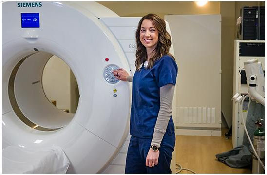
What will the scan be like?
First, you will be asked to lie on a table that slides inside a tunnel-shaped machine. The machine makes a lot of noise, so the technologist will give you earplugs to protect your hearing. The time it takes to scan will depend on what your doctor has ordered; generally, 30-45 minutes is to be expected. One of the most important things is that you hold very still once the scan has begun. Movement will blur or distort the pictures and lengthen the scan. If your test is ordered with contrast, the technologist will start an IV prior to scanning. Toward the end of the scan you will get an injection of contrast, then scanning will resume until all required images are taken. The contrast material used for MRI’s is less likely to cause an allergic reaction than the material used for CT scans.
What happens after the exam?
You may leave as soon as the scan is complete, unless you receive special instructions. You may resume normal activities as instructed by your doctor. It is recommended that you drink plenty of fluids to excrete any contrast from your system, if it was given. In the meantime, your scan will be prepared by the technologist to be examined and interpreted by the radiologist. A report of the radiologist’s findings will be sent to your doctor. You will need to make an appointment with your doctor to discuss the results. The MRI scan will help the doctor to plan appropriate treatment, if necessary.
What are the benefits associated with MRI?
There are many benefits to having an MRI. One major benefit is that the machine operates on magnetic fields and does not use radiation for generating images. They provide very detailed images in different planes so that the radiologist can get accurate measurements when evaluating your scan. They are generally painless and are a suitable scan for children and pregnant women when indicated. Safety studies have shown no long-term negative effects from MRI scans.
What are the disadvantages associated with MRI?
Disadvantages are minimal. Every MRI facility will have a screening procedure that will ensure the MRI is used on people for whom it is safe. If you have any metal within your body, you must inform the technologist. Many older medical implants can heat up tissues embedded in your body or dislodge wires, which can cause damage. MRI scans can cause heart pacemakers, defibrillation devices and cochlear implants to malfunction. Many newer medical implants are now manufactured to be MRI-compatible, so once the doctors know the exact nature of your implant they’ll be able to tell you if it’s safe for you to have an MRI. If you do have an implant that could make an MRI unsafe, the radiologist may recommend you have a different type of scan.
***If you have any additional questions or concerns, please give us a call, or talk with your physician.
*** Your physician has referred you for an MRI scan, a diagnostic procedure designed to reveal important information for use by your physician. He or she will be happy to tell you as much as you’d like to know about this important diagnostic scan. In the meantime, allow us to anticipate and answer some of the questions you’re likely to raise.

