
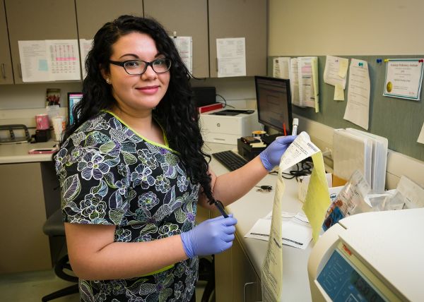


Diagnostic Imaging | PET/CT
What is a PET/CT (Positron Emission Tomography) scan and what are the benefits?
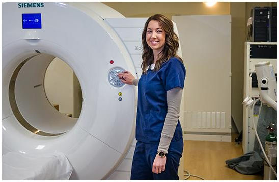
A PET/CT scan is a procedure that adds an important dimension to a physician’s ability to diagnose and manage disease. Our facility has a Siemens Biograph Horizon PET/CT machine that combines functional information from a PET scan with the anatomical information from a CT machine.
Instead of detecting changes in the physical size or structure of internal organs, as other imaging technologies, PET detects changes in cellular function. Since these functional changes may take place before physical changes occur, PET can often provide information that enables your physician to make an earlier diagnosis of diseases or abnormalities. If these diseases or abnormalities have already been detected by an imaging exam, such as CT or MRI study, PET can often characterize the cellular function early in the course of the disease.
These capabilities can, in turn, translate into faster initiation of the best possible treatment, often avoiding more invasive exams or exploratory surgery.
How will the scan be performed?
A PET/CT Technologist will explain the procedure and ask you about your medical history. After we have reviewed your history, we will start an I.V. in your arm and you will receive an injection of a radiopharmaceutical through your IV. You will then have to wait 45-60 minutes in a lounge chair for the radiopharmaceutical to distribute itself throughout your cells and organs.
Then, you will lie on a comfortable table that moves slowly through a ring-like PET scanner as it acquires the information it needs to generate diagnostic images. We will ask you to lie very still because movement can interfere with the results.
You should not feel a thing during the scan, which can last anywhere from 15 to 30 minutes. Then, unless there is a need for acquiring additional information, you will be free to leave. Afterward, you can go home and resume your normal activities.

